When to treat a Horseshoe tear?
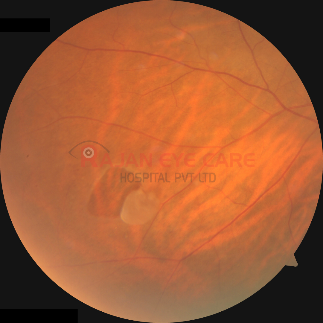
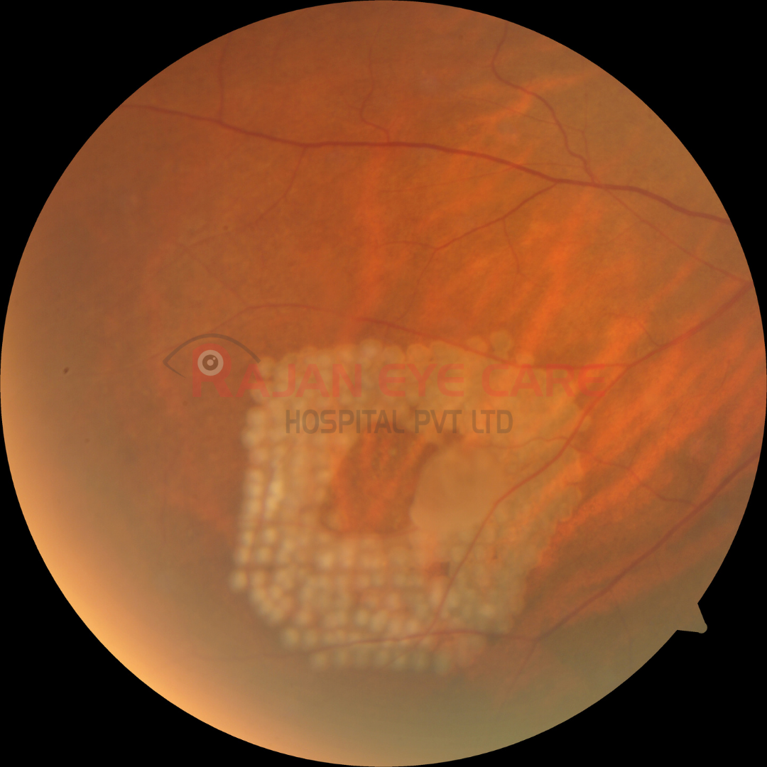
• The decision to treat a retinal tear is based on several factors, including symptomatology, patient age and systemic health, refractive error, location, age, type, size of the break; status of the fellow eye, lens status, and family history of retinal break and/or retinal detachment.
Symptomatic retinal tears
• Patients with symptomatic retinal horseshoe tears and persistent vitreoretinal traction can progress to a retinal detachment in 33 to 55 % of cases, and these patients should always be immediately treated.
• The goal of treatment is to create a chorioretinal adhesion around the tear to prevent vitreous fluid from entering the subretinal space and creating a retinal detachment.
• New onset floaters and flashes are the typical symptoms of an acute retinal break, and studies have shown that their presence is the most important prognostic criterion for progression to retinal detachment.
Asymptomatic retinal tears
• Approximately 5% of these patients will progress to a retinal detachment.
• Per the AAO Preferred Practice Patterns, asymptomatic horseshoe tears without subclinical RD 'often can be followed without treatment'.
• Prophylactic treatment in phakic patients that develop asymptomatic horseshoe tears without previous history of retinal disease or high myopia is controversial but often performed.
• Patients who have greater than 6D of myopia have an increased incidence of retinal detachment and the treatment of asymptomatic retinal tears in this group but is usually performed although still controversial.
• It is theorized that extensive prophylactic therapy has the potential to stimulate changes in the vitreous, which can result in increased subsequent vitreoretinal traction and resultant retinal tear and/or detachment.
• The management of asymptomatic horseshoe tears remains controversial in patients who need cataract surgery, in aphakic or pseudophakic patients, and in patients who have retinal detachments in their fellow eye.
• In general, because of the increased incidence of detachment in these scenarios, strong consideration should be given to prophylactic treatment.
Observation without treatment is indicated when:
• The tear is chronic
• The tear is demarcated by pigmentation
• There is absence of vitreo-retinal traction
• Found in selected asymptomatic patients
• In patients with a family history of retinal detachment, prophylactic therapy may be of value, especially in eyes with retinal breaks or visible retinal precursors of retinal tears.
• Laser photocoagulation treatment of retinal breaks utilizes argon, or an equivalent, laser.
• Two main delivery systems are used, the slit lamp and the indirect ophthalmoscope.
• In contrast to cryotherapy, chorioretinal adhesion seems to occur sooner, but maximal adhesion occurs roughly 7–10 days later.
• The retinal tear should be surrounded with a minimum of three concentric rows of laser.
• If the tear cannot be surrounded, laser or cryotherapy should extend to the ora serrata.
• Although the spots need not be confluent, there should be no more than half a spot size of untreated retina between burns.
• Settings are 200–500 mm spot size and 0.1–0.2 seconds application at the power necessary to generate a gray-white burn.
• In general, indirect ophthalmoscopic laser photocoagulation and trans-scleral retinal cryopexy and are preferred for anterior retinal breaks because of difficulty in treatment of the anterior margin at the slit lamp.
• Posterior breaks can be managed more easily with the slit lamp or an indirect laser delivery system.
Image from Rajan Eye Care Hospital
#ophthalmology #ophthal #doctor #health #medical #vision #education #optometry #medicalstudent #optometrist #medicine #eye #ophtho #ophthalmologist #ophthalmo #med #medicaleducation #ophthalmologyresident #ophthalmologyresidency #apaoyo #retina #horseshoetear #retinaldetachment #laserphotocoagulation
