Pseudophakic CME
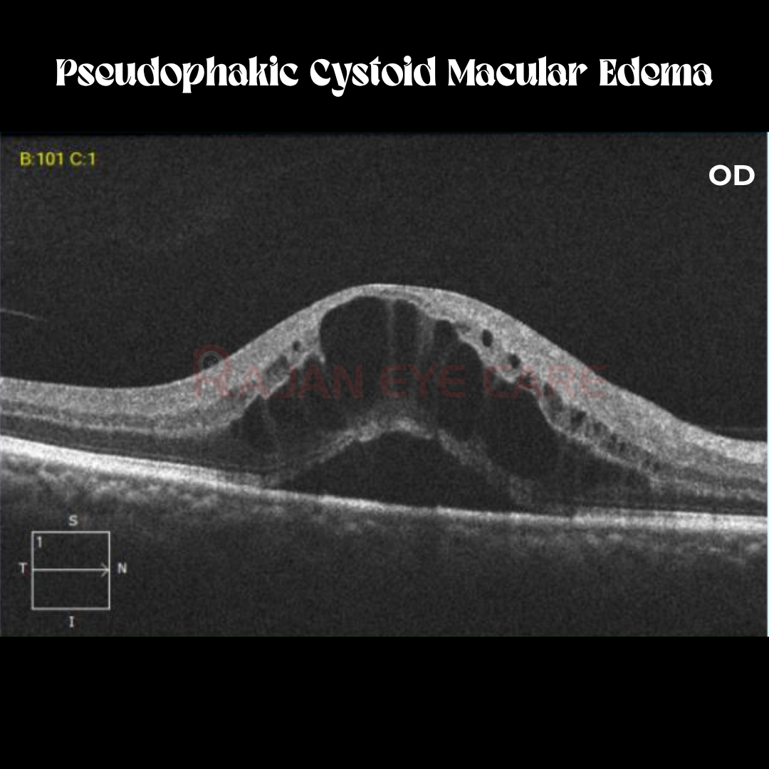
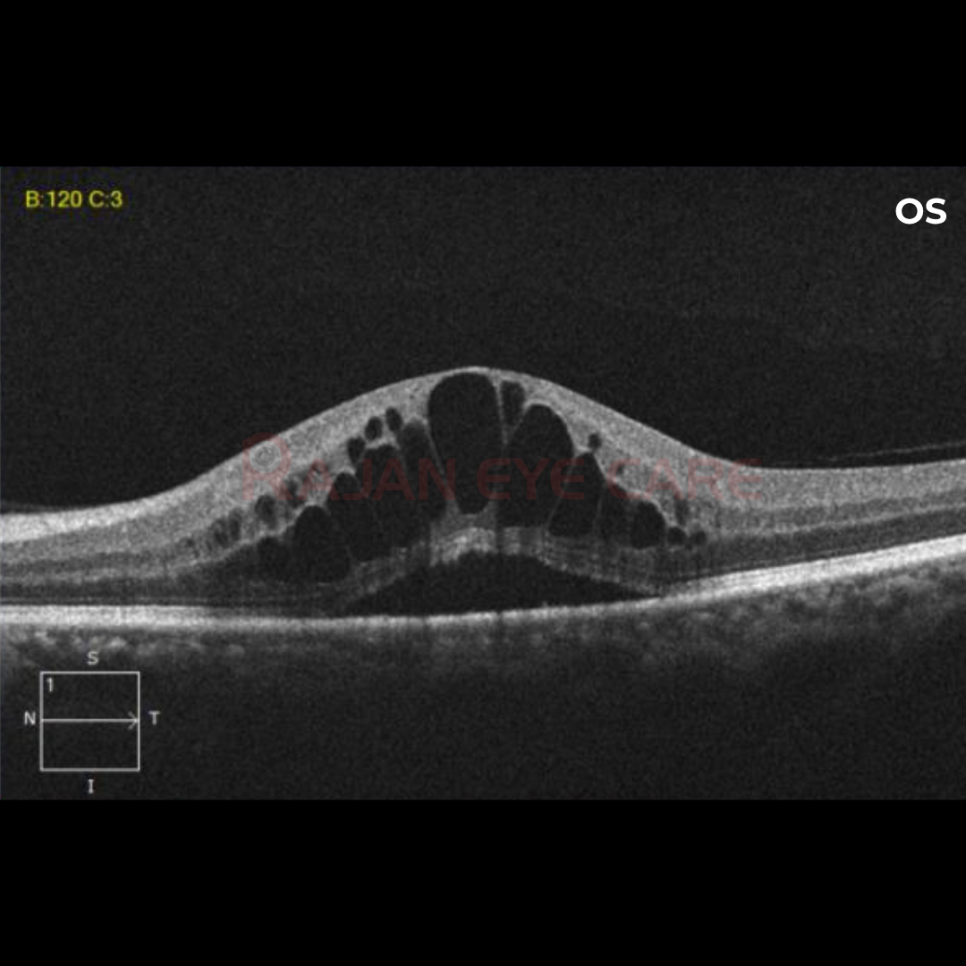
A 46 year old male diabetic came with complaints of DOV in both eyes 4 weeks after cataract surgery. There was a decrease in the near vision even with refractive correction. OCT macula was done and CME was confirmed
Pseudophakic cystoid macular edema (CME), also known as Irvine-Gass syndrome, is one of the most common causes of visual loss after cataract surgery
Pathogenesis
· The pathogenesis of pseudophakic CME is thought to be multifactorial.
· However, the major etiology appears to be inflammatory mediators that are upregulated in the aqueous and vitreous humors after surgical manipulation.
· Inflammation breaks down the blood-aqueous and blood-retinal-barriers, which leads to increased vascular permeability.
· Eosinophilic transudate accumulates in the outer plexiform and inner nuclear layers of the retina to create cystic spaces that coalesce to form larger pockets of fluid.
· In chronic CME, lamellar macular holes and subretinal fluid may also form
It's useful to distinguish:
· Angiographic CME — fluid leakage seen only on fluorescein angiography, often without symptoms
· Clinical CME — leakage accompanied by actual vision loss
· Acute CME — develops within about 6 months of surgery
· Chronic CME — persists beyond 6 months
Risk factors:
Surgical Complications
· Vitreous loss
· Vitreous traction at incision sites
· Vitrectomy for retained lens fragments
· Iris trauma
· Posterior capsule rupture
· Intraocular lens dislocation
· Early postoperative capsulotomy
· Iris-fixated intraocular lenses
· Anterior chamber intraocular lenses
Others
· DME
· Uveitis
· Glaucoma medication
History:
· The occurrence of pseudophakic CME peaks at approximately 4-6 weeks postoperatively. The most common presentation is blurry vision.
· Less common presentations include central scotomas, metamorphopsia, and mild photophobia.
· On biomicroscopy, retinal thickening and loss of the foveal depression is usually appreciated.
· In severe or chronic cases, optic disc swelling and/or a lamellar hole may also be seen. Splinter hemorrhages may also be present.
Imaging:
· FFA: characterized by retinal telangiectasis, capillary dilatation, and leakage from perifoveal capillaries in the early phase frames, and perifoveal hyperfluorescent spots classically described as a “petalloid” pattern in late phase frames, representing fluorescein accumulation in cystic spaces. Cystoid changes may also be apparent in the fovea and extramacular areas. Optic nerve staining is also commonly seen, and helps to distinguish pseudophakic CME from other causes of CME such as diabetic macular edema. FA is the gold standard in diagnosing pseudophakic CME, but treatment responses would be more conveniently monitored by biomicroscopy, visual acuity and OCT.
· OCT: Loss of the foveal depression, retinal thickening, and cystic hyporeflective areas within the macula.
· OCTA: disruption of parafoveal capillary arcade and cystoid spaces in the deep capillary plexus. Larger foveal avascular zone and reduced vessel density of full-thickness retina and DCP.
Management
Medical
· NSAIDs
· Corticosteroids: Topical/ periocular/ intravitreal
· Anti-VEGF
· Oral carbonic anhydrase inhibitors: in refractory CME
· Recent small pilot studies have started to examine subcutaneous interferon alpha and intravitreal infliximab
Surgical
· Neodymium:YAG laser anterior vitreolysis can release vitreous incarceration in the cataract incision wounds that complicate pseudophakic CME
· Pars plana vitrectomy may be considered when pseudophakic CME is complicated by vitreoretinal traction, and/or if the CME is unresponsive to other treatments
Image from Rajan Eye Care Hospital
#ophthalmology #ophthal #doctor #health #medical #vision #education #optometry #medicalstudent #optometrist #medicine #eye #ophtho #ophthalmologist #ophthalmo #med #medicaleducation #ophthalmologyresident #ophthalmologyresidency #apaoyo #pseudophakia #macularedema #cme #cystoidmacularedema #retina #macula
