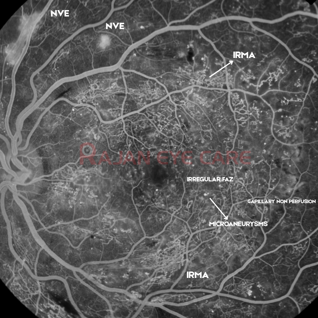
FFA of left eye in Arteriovenous to venous phase (Arteries and veins are fully filled, with no laminar flow left in the veins)
1. Neovascularisation: Large, fluffy, ill-defined hyperfluorescent patch lying along the vessels. Its fuzzy borders mean dye is leaking into the vitreous. New vessels lack tight junctions, so they leak heavily. That leakage is how FFA tells them apart from IRMA.
Round fluffy patch above the superotemporal arcade (upper centre-left). It looks like a smaller NVE frond.
2. IRMA (intraretinal microvascular abnormalities): Clusters of tortuous, dilated, beaded, capillary segments, densest above the macula, temporal to the fovea, and in the inferior arcade region.
IRMA are shunt vessels at the edges of non-perfused retina. They stay within the retina and leak only minimally, unlike NVE.
Extensive IRMA in all quadrants is a severe ischaemic sign in itself.
3. Capillary non-perfusion (ischaemia):
There are dark, featureless zones where the fine capillary network is missing. These lie between the arcades, in the temporal macula, and towards the periphery.
The foveal avascular zone looks enlarged and irregular, which suggests macular ischaemia. That carries a guarded visual prognosis even after treatment.
Widespread non-perfusion drives VEGF release, which in turn produces the neovascularisation.
4. Microaneurysms:
Many pinpoint hyperfluorescent dots are scattered across the posterior pole. They fill early and may leak late.
5. Other points
The veins look dilated and slightly irregular. Look for venous beading on closer frames.
There is mild diffuse hyperfluorescence around the fovea. In late phase this could be diabetic macular oedema, and OCT is the best way to confirm it.
This is untreated proliferative diabetic retinopathy. It shows leaking NVE, extensive IRMA, widespread capillary non-perfusion including an enlarged FAZ, microaneurysms, and blocking haemorrhages.
Management implications:
Panretinal photocoagulation and/or intravitreal anti-VEGF.
OCT to check for DMO.
Systemic control of HbA1c, blood pressure and lipids.
Close follow-up, given the risk of vitreous haemorrhage and tractional detachment.
Image from Rajan Eye Care Hospital
Visit our website www.ophthalmobytes.com
#ophthalmology #ophthal #doctor #health #medical #vision #education #optometry #medicalstudent #optometrist #medicine #eye #ophtho #ophthalmologist #ophthalmo #med #medicaleducation #ophthalmologyresident #ophthalmologyresidency #apaoyo #ffa #retina #diabeticretinopathy #pdr #proliferativediabeticretinopathy
