Post Trab: How to assess?
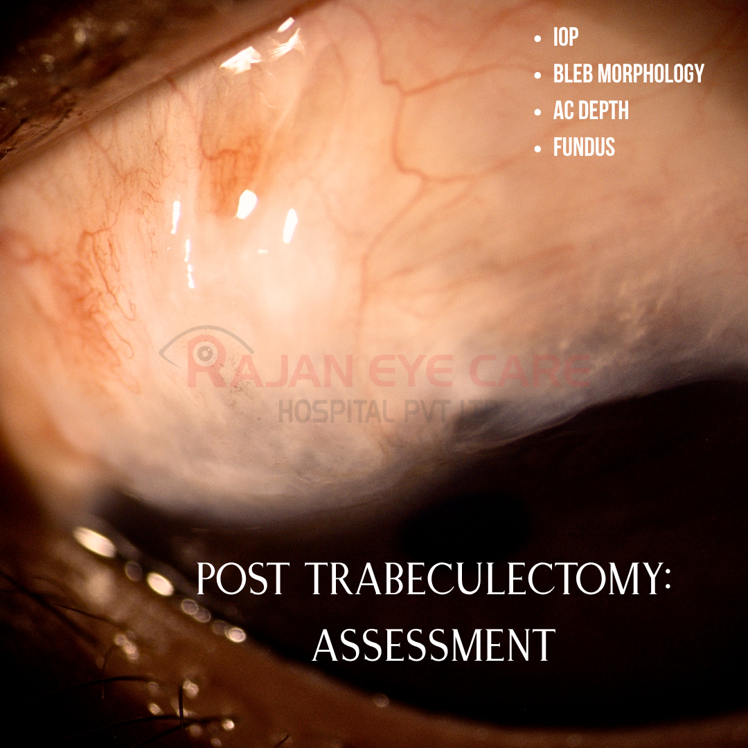
•Post-trabeculectomy assessment focuses on monitoring IOP, examining filtering bleb morphology, checking for leaks (Seidel test), and maintaining ocular surface health to ensure surgical success
•Early, frequent follow-ups are critical for titrating medications and adjusting sutures to maintain target IOP
What to look for?
1) IOP Monitoring
•Early (first days/weeks) control is crucial to determine long-term success, as low IOP can cause complications like choroidal detachment, while high IOP indicates failure
•Target IOP between 8-12mmHg depending on the severity of glaucoma
2) Bleb Evaluation
•Slit-lamp exam for bleb morphology (height, vascularity, spread) and functionality
•Morphology: Assessed for height (flattened, low, medium, high), extent (diffuse vs. localized), and location
•Vascularization: Lower vascularity indicates better function
•Microcysts: Presence in the conjunctival epithelium is a positive sign
•Seidel's Test: Assesses for aqueous leakage (negative is generally preferred, although some flow is expected early on)
•AS-OCT: to evaluate maximal height, bleb wall thickness and subconjunctival spaces
Moorfields Bleb Grading system
•Diffuse: Thin-walled blebs with large surface area and low elevation
•Microcystic: Thin-willed blebs on the scleral bed with moderate elevation, large surface area and microcystic structure
•Flat: No important signs of bleb development such as elevation or microcysts
•Encapsulated: Thick-walled blebs with cystic appearance, high elevation and demarcated area
How to describe a bleb?
•Elevation
•Vascularisation
•Thickness of wall
•Extent in clock hours
•Localized/ diffuse
•Presence/ absence of microcysts
•Functionality
•Diffuse appearance, slight elevation above the scleral flap, the presence of microcysts in the conjunctival epithelium and low conjunctival vascularization indicate bleb functionality, while a narrow surface area, excessive elevation and dense vascularization are indicators of bleb failure
3) Anterior Chamber Depth: •Monitoring for shallow AC, related to over-filtration or leak
•Look for hyphema
4) Conjunctival & Wound Integrity:
•Checking for proper healing of the conjunctival sutures and the absence of infection or excessive fibrosis (scarring)
5) Ophthalmoscopy
•Assessing the ONH and monitoring for complications like hypotony maculopathy, choroidal detachment
Image from Rajan Eye Care Hospital
#ophthalmology #ophthal #doctor #health #medical #vision #education #optometry #medicalstudent #optometrist #medicine #eye #ophtho #ophthalmologist #ophthalmo #med #medicaleducation #ophthalmologyresident #ophthalmologyresidency #apaoyo #glaucoma #bleb #trabeculectomy #iop #opticnerve