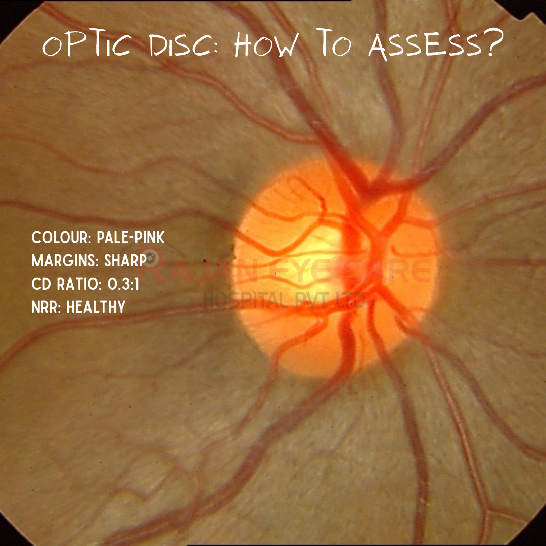
Optic Disc: How to assess?
Key Features of a Normal Optic Nerve
-Color: Healthy pink, indicating good vascularity; not pale or white.
-Border/Margin: Sharp and well-defined.
-Cup-to-Disc Ratio (CDR): Generally 0.3 or less, meaning the pale central depression occupies less than one-third of the total disc diameter
-Neuroretinal Rim: The tissue surrounding the cup should be thick and pink.
-ISNT Rule: The thickness of the rim usually follows the pattern: Inferior>Superior>Nasal>Temporal.
-Surface: Flat, not significantly elevated or depressed.
-Vessels: No hemorrhage, abnormal engorgement, or vessel displacement.
DISC SIZE:
-Cup size is linearly related to disc size.
Clinical Implications:
-Large discs → large physiologic cups (can look glaucomatous but are normal)
-Small discs → small cups; even mild cupping may be significant
Missing disc size assessment leads to:
-Overdiagnosis in large discs
-Missed glaucoma in small discs
Measurement Tips:
-90D lens:
-Average: ~1.4 mm
-Large: >1.6 mm
-Small: <1.2 mm
The SHIP Framework
S – Size
H – Hemorrhages
I – ISNT rule violation
P – Parapapillary atrophy
Types of Glaucomatous Rim Loss
1. Generalized Concentric Loss
2. Focal Rim Loss
3. Mixed (Most common)
4. Senile Sclerotic Disc
5. Myopic Tilted Discs
Parapapillary Atrophy (β-PPA)
-Often an early sign
-Increases with progression
-Important in small discs
-Frequently accompanies focal loss
RNFL Changes:
-Widening nerve fiber layer defects
-Precede visual field loss
-Often associated with rim thinning and vessel shifts
PALLOR
-A Warning Sign
-May accompany cupping
-If pallor > cupping → suspect non-glaucomatous optic neuropathy
-Can follow acute IOP spikes
ADVANCED GLAUCOMA SIGNS
-Deep excavation
-Exposure of scleral ring of Elschnig
-Severe rim loss
-Vessel baring
How to Detect Progression
Look for:
-Thinning rim
-Widening notch
-Disc hemorrhage
-Vessel shifts
-Increased β-PPA
-Pallor
-Widening RNFL defects
When you see an optic nerve:
1) Assess size first
2) Apply SHIP
3) Look for focal rim thinning
4) Search for disc hemorrhage
5) Examine RNFL
6) Compare with prior photos
7) Decide: Stable or progressing?
Image from Rajan Eye Care Hospital
#ophthalmology #ophthal #doctor #health #medical #vision #education #optometry #medicalstudent #optometrist #medicine #eye #ophtho #ophthalmologist #ophthalmo #med #medicaleducation #ophthalmologyresident #ophthalmologyresidency #apaoyo #opticdisc #glaucoma #disc #opticnerve