Optic Disc Signs
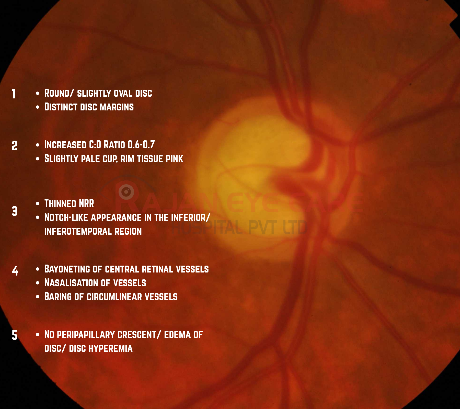
•The main abnormality is an enlarged and somewhat irregular optic cup with focal rim thinning (notching) and vessel bayoneting
Vascular changes seen on the optic disc are:
•Nasalisation of Vessels
•Bayoneting
•Overpass vessels
•Narrowing of retinal arterioles
•Baring of Circumlinear Vessels
•Collaterals
Bayoneting:
•In areas where the neuroretinal rim is thin or absent, a retinal vessel may pass under the overhanging edge of the cup and make a sharp bend as this crosses the cup margin.
•It is due to the neural tissue loss extending below the edge and is named so because of its resemblance to the sharp angle of a rifle bayonet.
Overpass vessels:
•Normally, the retinal vessels crossing the disc margin pass on or through the surface of the neuroretinal rim.
•If the underlying rim is lost, the vessel may appear to “hang in the air” without any contact with the underlying tissue
Baring of Circumlinear Vessels
•A circumlinear vessel is a small branch of the central retinal vein or artery following a curved path along the optic cup margin.
•When the cup enlarges, the margin recedes from the vessel leaving a space between the margin and the vessel
•Often present in glaucomatous discs.
Image from Rajan Eye Care Hospital
#ophthalmology #ophthal #doctor #health #medical #vision #education #optometry #medicalstudent #optometrist #medicine #eye #ophtho #ophthalmologist #ophthalmo #med #medicaleducation #ophthalmologyresident #ophthalmologyresidency #apaoyo #opticdisc #glaucoma #glaucomatousopticatrophy #opticdiscsigns #retina
