How do we assess a corneal tear wound?
Before assessment of wound,
-A quick assessment of the visual acuity of the injured eye must be performed
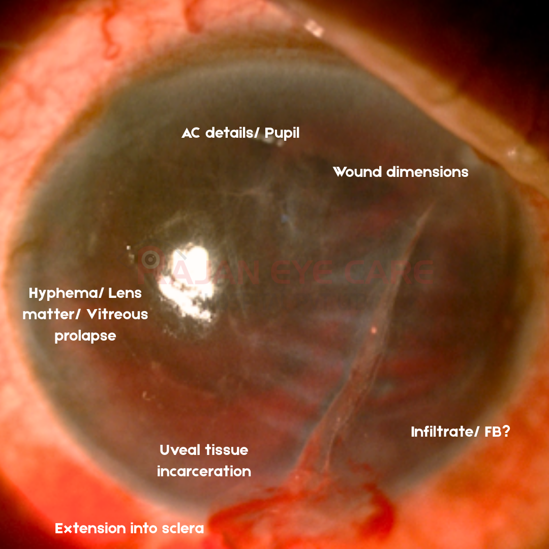
-Location of wound whether involving the visual axis can give us an idea on the visual prognosis of the eye
-Length and depth of wound must be documented as well as any extension into scleral tissue
-The presence of foreign bodies, any uveal tissue incarceration, lens rupture, lens material in AC, vitreous prolapse and the presence of or potential for infection must be determined
-History can give us a clue about suspected foreign body in the patients eye
-In case of suspected foreign body, Xray or CT imaging needs to be done
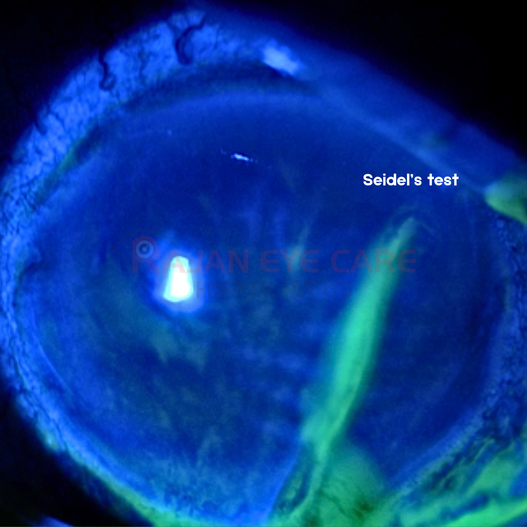
-A Seidel’s test can be done for checking for leak in suspect cases
-Explain the visual prognosis and obtain a consent
-The status of tetanus prophylaxis of the individual must be ascertained in the event of a surgical repair being considered
-Once the assessment is complete, the eye must be patched with a rigid shield and patient is also instructed to avoid straining further
-General anaesthesia is preferred in these patients to prevent upthrust during surgery
-B scan must be deferred till post operative period in open globe injuries
www.ophthalmobytes.com
Image from Rajan Eye Care Hospital
#ophthalmology #ophthal #doctor #health #medical #vision #education #optometry #medicalstudent #optometrist #medicine #eye #ophtho #ophthalmologist #ophthalmo #med #medicaleducation #ophthalmologyresident #ophthalmologyresidency #apaoyo #cornea #cornealtrauma #trauma #cornealtear #openglobe #seidelstest #cornealtearrepair