Acute Corneal Hydrops
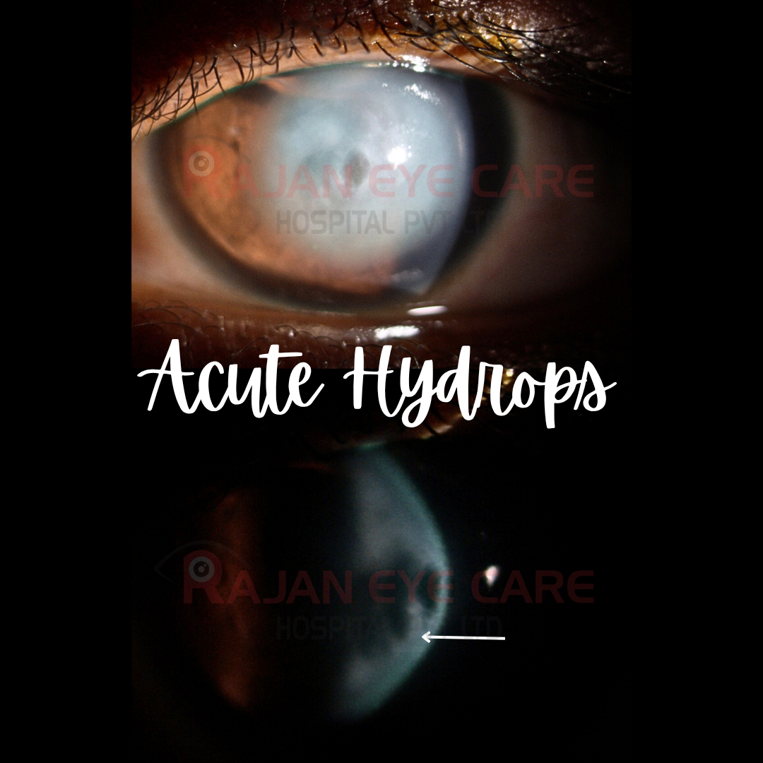
•Development of marked corneal edema due to a tear in the DM & posterior stroma followed by leakage of aqueous into stroma
RISK FACTORS:
•Allergic eye disease
•Progressive ectasia
•Eye rubbing
•Down syndrome
•Steep K
PATHOGENESIS:
•It results from a break in Descemet membrane, Pre-Descemet's and the endothelium, leading to an influx of aqueous humor into the stroma and subsequent formation of corneal edema
•Continuous accumulation of the aqueous leads to the separation of the collagen lamellae and the formation of large fluid-filled stromal pockets
PREVENTION:
•Controlling the progression of corneal ectatic disorders can lower the risk
•Minimizing risk factors such as eye rubbing and limiting exposure to allergens
DIAGNOSIS:
1) Slit-lamp
2) AS-OCT: allows for visualization of the rupture in DM or posterior stroma, and the extent of detachment from the cornea
3) IVCM: demonstrates epithelial and stromal edema
4) UBM: assessment of corneal edema and intrastromal clefts
5) Corneal Tomography: of the fellow eye
MANAGEMENT:
MEDICAL:
•Hypertonic saline
•Topical steroids
•Cycloplegics
SURGICAL:
•Air/gas injection
•Compression sutures
•DALK
•PK
SEQUELAE:
•Development of vision-debilitating scar
Image from Rajan Eye Care Hospital
#ophthalmology #ophthal #doctor #health #medical #vision #education #optometry #medicalstudent #optometrist #medicine #eye #ophtho #ophthalmologist #ophthalmo #med #medicaleducation #ophthalmologyresident #ophthalmologyresidency #apaoyo #cornea #keratoconus #hydrops