What are the differences between the central cornea and peripheral cornea?
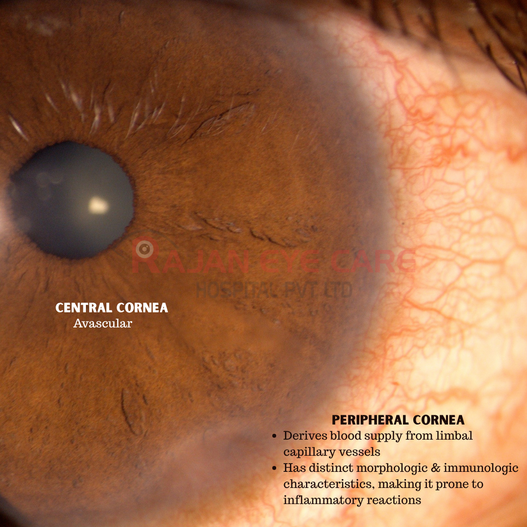
• Thickness: The central cornea is thinner (about 0.5 mm), while the peripheral cornea is thicker (about 1.0 mm)
• Shape: The center is steeper, whereas the periphery is flatter
• Nerves and Sensitivity: The central area has significantly more nerve fibers, making it much more sensitive to touch than the periphery
• Cell organization: Central cells and collagen fibers are tightly, precisely arranged for vision, while peripheral fibers transition into a looser, wider structure with larger collagen fibril diameters
• Vascularity: The central cornea contains no blood vessels to maintain perfect clarity. The peripheral cornea is closer to limbal conjunctiva and derives its blood supply and lymphatics from the limbal capillary vessels, which extend approximately 0.5 mm into the clear cornea
• This is a source of immunocompetent cells, such as macrophages, Langerhan’s cells, lymphocytes, and plasma cells
Immune complexes circulating in the blood of the patients with collagen vascular disease can lodge at the ends of the limbal vessels by virtue of their size and the restrictive molecular sieving characteristics of the cornea
• The peripheral cornea contains five times higher levels of the first component of the complement (C1) than does the central cornea which stimulates chemotaxis of the neutrophils to the area
• The concentration of IgM is greater at this site
Immune complexes of rheumatoid factor and Ig G can deposit in the peripheral cornea and produce inflammation and corneal ulceration
• Langerhans’ cells, which are the pre-eminent antigen-presenting cell, are also concentrated more densely in the peripheral cornea
Image from Rajan Eye Care Hospital
#ophthalmology #ophthal #doctor #health #medical #vision #education #optometry #medicalstudent #optometrist #medicine #eye #ophtho #ophthalmologist #ophthalmo #med #medicaleducation #ophthalmologyresident #ophthalmologyresidency #apaoyo #cornea #centralcornea #peripheralcornea
