Nodules to Neuro!
𝐋𝐢𝐬𝐜𝐡 𝐍𝐨𝐝𝐮𝐥𝐞𝐬
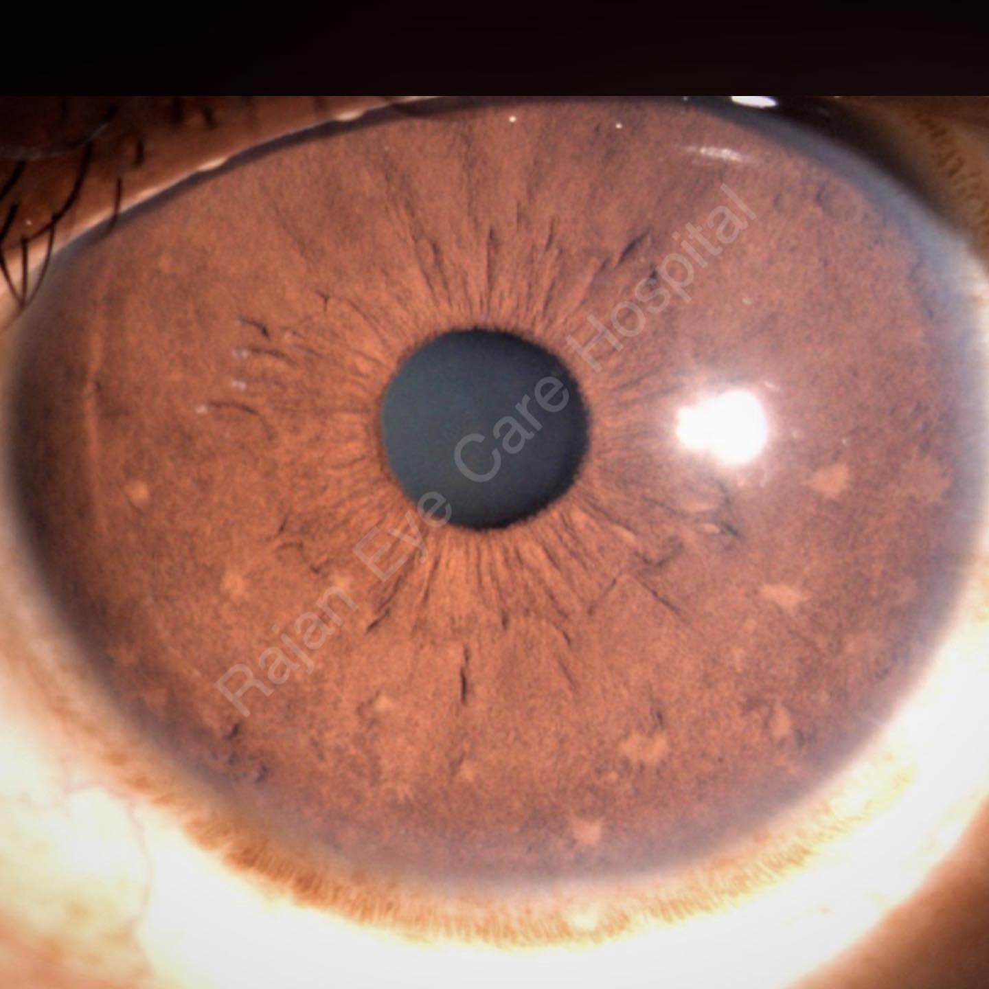
Melanocytic hamartoma of iris
Associated with Neurofibromatosis 1 (Autosomal dominant with full penetrance) - multiple and bilateral
𝘋𝘦𝘴𝘤𝘳𝘪𝘱𝘵𝘪𝘰𝘯
● Well-defined, dome shaped elevations, tan - yellow brown coloured, projecting from the surface of the iris
● Best identified by slit lamp examination
● Visible in children after the age of 6 years
● Size and number increase with age (100% over 21 years of age)
● Important to differentiate them from nevi which are flat/ minimally elevated, densely pigmented, blurred margins
● Multiple Lisch nodules point to the diagnosis of NF 1
𝘏𝘪𝘴𝘵𝘰𝘭𝘰𝘨𝘺
Closely packed dendritic or spindle shaped melanocytes within the anterior layers of iris stroma - normal uveal melanocytes and not nevus cells
Diagnostic Criteria of NF 1
1. Six or more cafe-au-lait macules over 5 mm in greatest diameter in prepubertal individuals and over 15 mm in greatest diameter in postpubertal individuals
2. Two or more neurofibromas of any type or one plexiform neurofibroma
3. Freckling in the axillary or inguinal region
4. Optic glioma
5. Two or more Lisch Nodules (iris hamartomas)
6. A distinctive osseous lesion such as sphenoid dysplasia or thinning of long bone cortex, with or without pseudarthrosis
7. A first-degree relative (parent, sibling, or offspring) with neurofibromatosis type 1 by the above criteria.
Image from Rajan Eye Care Hospital
#ophthalmology #ophthal #irisnodules #iris #neurofibromatosis #lischnodules #systemicophthalmology