5 features of Grade 4 Hypertensive Retinopathy
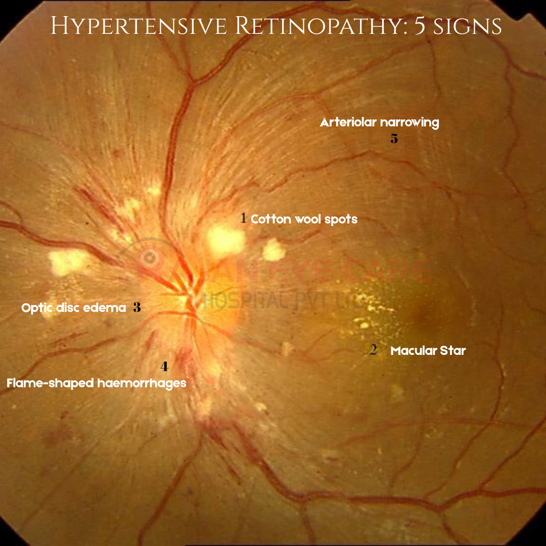
1) Cotton wool spots
•Multiple soft white patches near the disc representing nerve fiber layer infarcts due to ischemia.
2) Hard exudates forming a macular star
•Yellow lipid deposits arranged in a star-shaped pattern around the macula
•This occurs because exudates spread along the Henle fiber layer
3) Optic disc edema (papilledema)
•The optic disc appears hyperemic and swollen with blurred margins
•There are radial peripapillary exudates and congestion of vessels around the disc
4) Flame-shaped hemorrhages
•Seen around the optic disc and along the nerve fiber layer
•These are typical superficial retinal hemorrhages caused by rupture of precapillary arterioles under high pressure
5) Arteriolar narrowing and vascular changes
•Generalized arteriolar narrowing with increased vascular tortuosity
This corresponds to Keith–Wagener–Barker Grade IV Hypertensive Retinopathy
Grade IV features include:
•Retinal hemorrhages
•Cotton wool spots
•Hard exudates (often macular star)
•Papilledema
CLINICAL SIGNIFICANCE
•Indicates malignant hypertension
•Associated with hypertensive emergency
May be accompanied by:
•Renal failure
•Encephalopathy
•Cardiac failure
MANAGEMENT:
•Requires urgent blood pressure control
Image from Rajan Eye Care Hospital
Visit our website www.ophthalmobytes.com
#ophthalmology #ophthal #doctor #health #medical #vision #education #optometry #medicalstudent #optometrist #medicine #eye #ophtho #ophthalmologist #ophthalmo #med #medicaleducation #ophthalmologyresident #ophthalmologyresidency #apaoyo #opticdisc #retina #fundus #hypertensiveretinopathy #papilledema