Drance Hemorrhages
𝘖𝘱𝘵𝘪𝘤 𝘋𝘪𝘴𝘤 𝘏𝘦𝘮𝘰𝘳𝘳𝘩𝘢𝘨𝘦𝘴
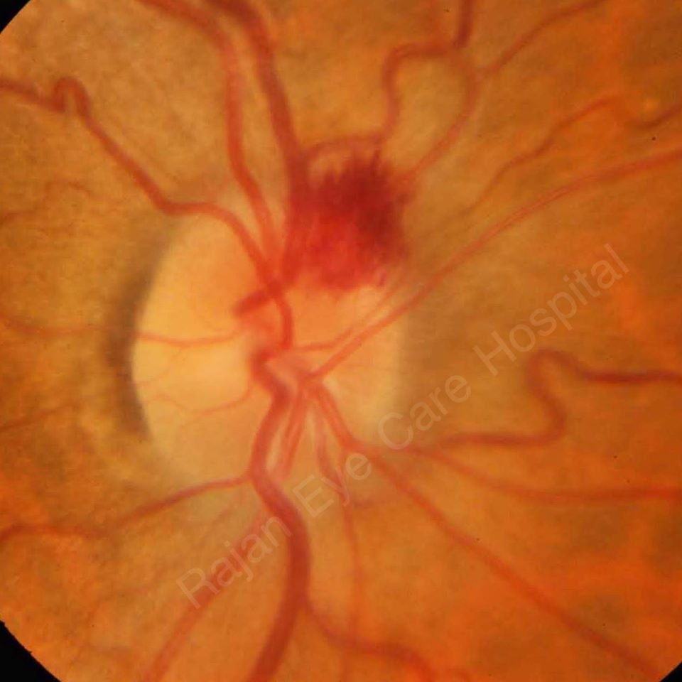
● Splinter or flame-shaped hemorrhages
● Perpendicular to the optic disc margin
● Classically, these hemorrhages are located in the prelaminar optic disc, cross the peripapillary zone, and extend into the adjacent superficial retinal nerve fiber layer
● Disc hemorrhages are a risk factor for progression of glaucoma
● Most common in normal tension glaucoma
● Associated with notching of the neuroretinal rim or retinal nerve fibre layer defects
𝐏𝐚𝐭𝐡𝐨𝐠𝐞𝐧𝐞𝐬𝐢𝐬:
𝘔𝘦𝘤𝘩𝘢𝘯𝘪𝘤𝘢𝘭 𝘛𝘩𝘦𝘰𝘳𝘺: .
● Disc hemorrhages result from mechanical shearing at the lamina cribrosa or because of damage to the capillary network at the border of retinal nerve fibre layer defect enlargement.
● Primary insult is neurodegenerative, and the hemorrhage is a secondary event resulting from tissue damage.
𝘝𝘢𝘴𝘤𝘶𝘭𝘢𝘳 𝘛𝘩𝘦𝘰𝘳𝘺:
● Ischemic microinfarction in the optic nerve head or break down of the blood-retinal barrier.
𝘓𝘰𝘤𝘢𝘵𝘪𝘰𝘯: Disc hemorrhages associated with glaucoma are found mostly in the inferotemporal and superotemporal regions of the optic disc
𝘚𝘺𝘴𝘵𝘦𝘮𝘪𝘤 𝘢𝘴𝘴𝘰𝘤𝘪𝘢𝘵𝘪𝘰𝘯𝘴:
● Hypertension
● Vascular disease
● Migraine
● Diabetes
● Aspirin use
● Smoking
● Exfoliation syndrome etc.
𝘛𝘳𝘦𝘢𝘵𝘮𝘦𝘯𝘵:
A disc hemorrhage does not need to be treated but its presence may signal the need for better control of intraocular pressure in glaucomatous eyes.
Image from Rajan Eye Care Hospital
#ophthalmology #ophthal #doctor #health #medical #vision #education #optometry #medicalstudent #optometrist #medicine #eye #ophtho #ophthalmologist #ophthalmo #med #medicaleducation #ophthalmologyresident #ophthalmologyresidency #glaucoma #opticdisc #opticdischemorrhage #disc #retina