Epithelial Ingrowth after LASIK
Epithelial cells invade the space between the corneal flap and underlying stromal bed.
ETIOLOGY:
-Unclear etiology, however, poor flap adhesion or apposition may allow surface epithelium to migrate into this space. -Disturbance of the corneal epithelium intraoperatively or post-operatively
-Associated with flap melt
RISK FACTORS:
Preoperative:
-EBMD
-History of RCE
-Increased patient age
-Diabetes mellitus
-Epithelial ingrowth in the contralateral eye
Operative:
-Intraoperative epithelial defect
-Postoperative inflammation (lamellar keratitis)
-Flap relift, enhancement procedure
-Flap edema from any cause, flap misalignment or shift
-Ablation extending past the flap diameter
-Irregular flaps
-Buttonholes
-Free cap
PATHOLOGY:
Stratified squamous epithelial cells with a basement membrane invade the intrastromal interface made during the LASIK procedure
PRIMARY PREVENTION
-LASIK patients should be evaluated for risk factors which may lead to an intraoperative epithelial defect
-The surgeon should practice excellent surgical technique with minimal epithelial manipulation and accurate flap apposition
-Excessive topical anesthetic use, interface irrigation, or surface drying may contribute to epithelial defect formation
-BCL should be strongly considered in the event of an intraoperative epithelial defect. -
When performing an enhancement procedure, attention should be given to removing the peripheral epithelium from the flap interface and obtaining excellent flap apposition when replacing the flap.
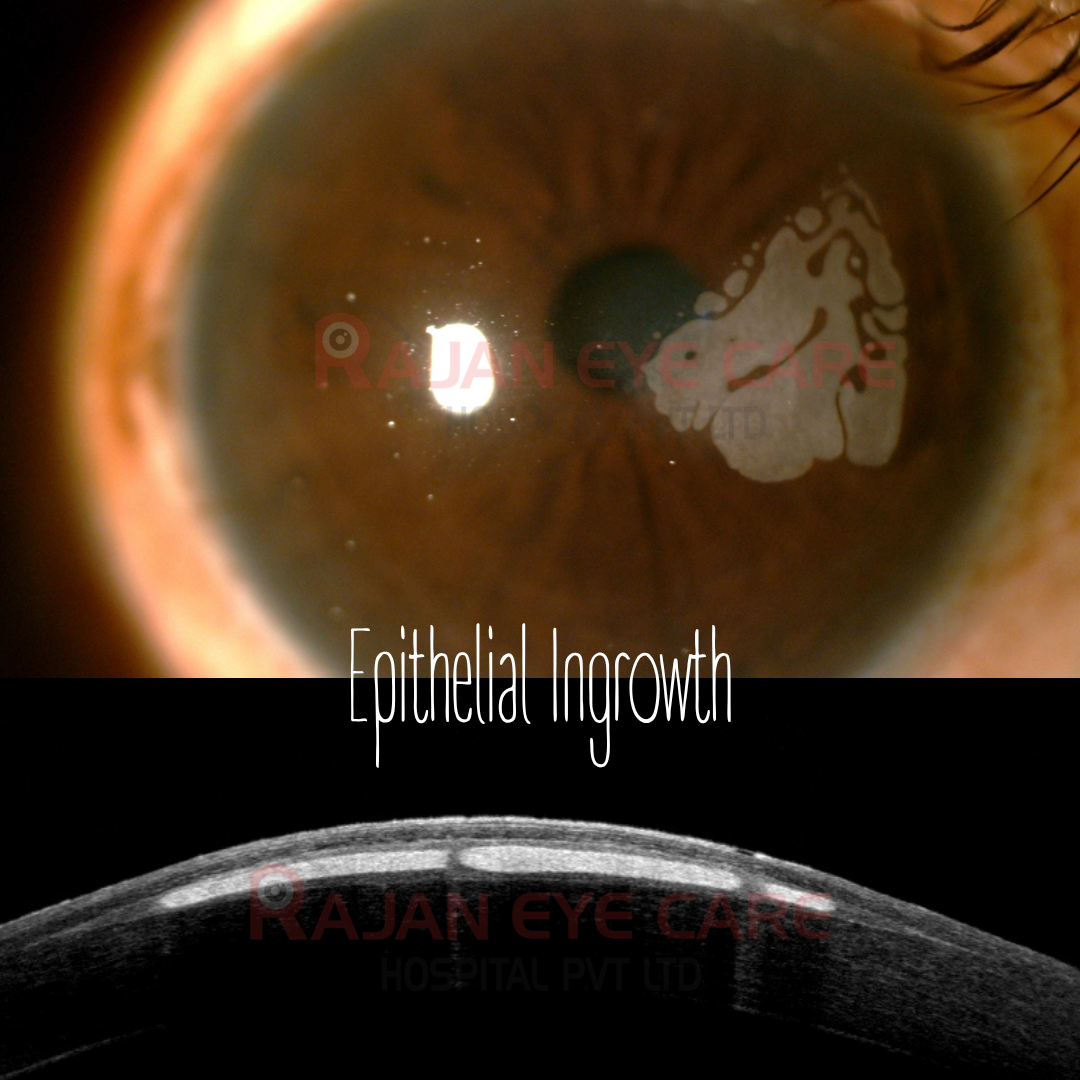
SIGNS:
-Nests of cells at flap interface, usually close to the edge of the flap
-Fibrosis in area of longstanding epithelial ingrowth
-White plaque of cells in flap interface
-Irregular astigmatism
-Fluorecein staining at border of flap where cellular invasion may originate
CLASSIFICATION:
Probst/Machat epithelial ingrowth classification:
GRADE 1: Thin, localized nests of epithelial cells confined to an area within 2.0 mm of the flap edge. (No treatment)
GRADE 2: Thicker growth that is slowly progressive but remains within 2.0 mm of the flap edge. (Requires non-urgent treatment within 2-3 weeks)
GRADE 3: Pronounced, progressive, and opaque growth that has extended beyond 2.0 mm from the flap edge. (Urgent treatment required with close follow-up due to frequent recurrences)
MANAGEMENT:
-Removing the invading epithelial cells from the interface and achieving closure of the flap edge to prevent recurrent invasion of epithelium into the flap stromal interface space
-Adjuvant treatments such as ethanol, mitomycin, PTK have been described for recurrent epithelial ingrowth, but may cause adverse effects
Image from Rajan Eye Care Hospital
#ophthalmology #ophthal #doctor #health #medical #vision #education #optometry #medicalstudent #optometrist #medicine #eye #ophtho #ophthalmologist #ophthalmo #med #medicaleducation #ophthalmologyresident #ophthalmologyresidency #apaoyo #cornea #lasik #refractivesurgery #femtolasik #epithelialingrowth #asoct