Basal Cell Carcinoma of Eyelid
-Constitutes 85-90% of all malignant eyelid epithelial tumours.
MECHANISM:
-Arises from a pluripotent stem cell in the epidermis that proliferates, amplifies and terminally differentiates
-Enhanced tumour cell motility and collagenase content
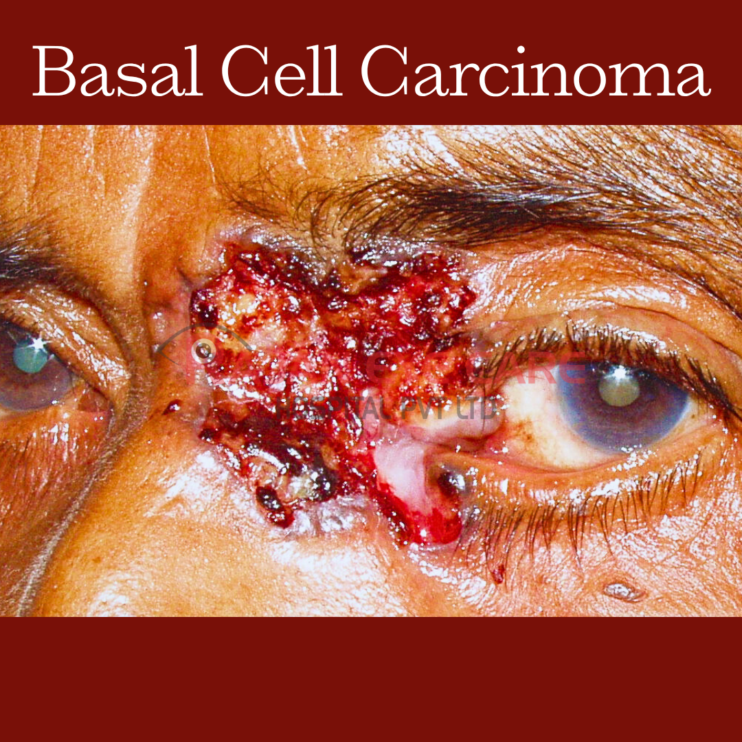
LOCATION:
-50-60% affect lower eyelid
Medial canthus: 25-30%
Upper lid 15%
Lateral canthus 5%
TYPES:
1) Nodular-ulcerative:
-Most common
-Pink/pearly papule or nodule with overlying telangiectatic vessels
-Central ulceration with rolled border.
-Called rodent ulcer
2) Pigmented
-Similar to nodular-ulcerative in morphology but with brown/black pigmentation
-Resemble malignant melanoma.
3) Morphea or Sclerosing
-Flat, indurated, yellow-pink plaque with ill-defined borders
-May stimulate blepharitis or dermatitis
-Aggressive
-Invades dermis deeply
-Occurs in medial canthal region
-May invade into PNS and orbit.
4) Superficial
-Erythematous, scaling patch with raised pearly border
-Arise on the trunk.
5) Fibroepithelioma
-Pedunculated or sessile, smooth, pink nodule
-Arise on the trunk.
SYSTEMIC ASSOCIATIONS:
-Basal cell nevus syndrome (Gorlin-Gotz syndrome)
-Palmar and plantar pits
-Bazex syndrome
-Linear unilateral basal cell nevus
-Rombo syndrome
-Albinism.
-Xeroderma pigmentosa.
-Nevus sebaceus.
TREATMENT:
-Moh's Micrographic Surgery
-Excisional biopsy with frozen section control
-Radiation therapy (CI in basal cell nevus syndrome)
-Cryotherapy (outside periorbital area)
-Chemotherapy: 5% imiquimod
-Photodynamic therapy
www.ophthalmobytes.com
Image from Rajan Eye Care Hospital
#ophthalmology #ophthal #doctor #health #medical #vision #education #optometry #medicalstudent #optometrist #medicine #eye #ophtho #ophthalmologist #ophthalmo #med #medicaleducation #ophthalmologyresident #ophthalmologyresidency #apaoyo #eyelid #lid #basalcellcarcinoma #bcc #oculoplasty #ocularoncology