Paton’s Lines
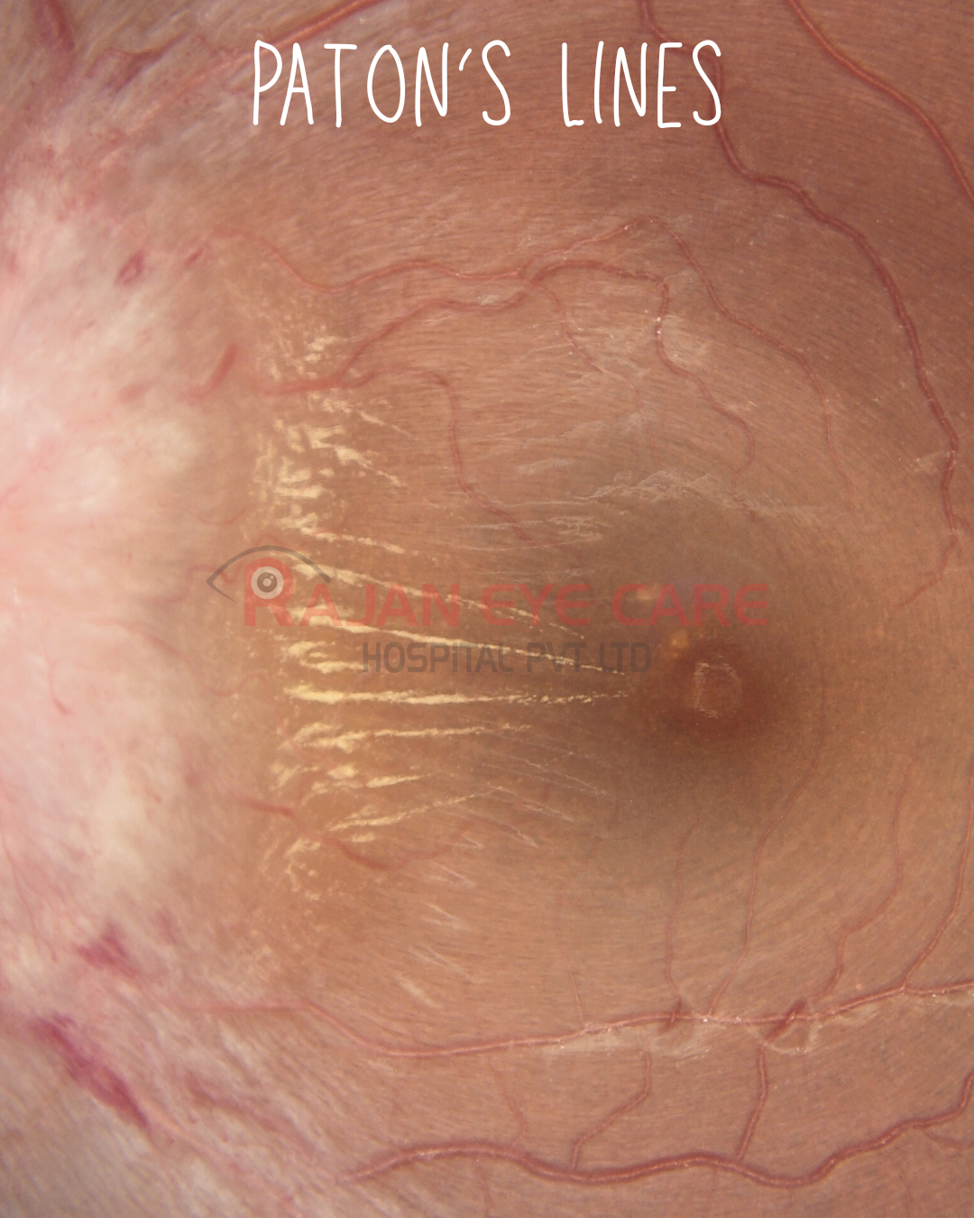
-Ophthalmoscopic finding of radiating lines around the optic disc
-Includes a structural display of tensile, compressive, or shearing stress that causes mechanical strain from pressure changes arising from pathological optic disc edema
-The disc edema dislodges surrounding structures to cause bulging, which appears as lines or folds
ETIOLOGY
-Most common cause: papilledema from intracranial hypertension
-Other causes of optic disc edema
-Non-arteritic anterior ischemic optic neuropathy
-Some maculopathies
-Orbital and intraocular tumors
-Orbital, choroidal, or scleral infiltrative and inflammatory diseases
-Ocular hypotony
-Retinal detachment
-Scleral buckles
-Epiretinal membranes
-Microgravity space flight associated neuro-ocular syndrome
-OCT can identify and differentiate the various types of ophthalmoscopically visible lines in the fundus including peripapillary wrinkles, retinal folds, choroidal folds and the various entities referred to in the pre-OCT era as Paton's lines
Clarifying the specific location of the folds can help determine the etiology of the pathological disc edema.
PATHOLOGY
-They may be found in the retina, choroid, or in the retina nerve fiber layer
-PL in the retina are either intraretinal or on the surface
-3 distinct ophthalmoscopic patterns of PL in the retina have been described including concentric, spiral, or radial lines
-RF, CF or PPW can be found exclusively or concurrently with each other
-These patterns are associated with distinct forms of stress
RETINAL FOLDS
-Concentric, spiral, and radial patterns of RF are located in or on the retina
-The most specific pattern of RF due to papilledema is radial RF
CHOROIDAL FOLDS
-CF are undulations of the choroid, which in contrast to retinal folds, are located deeper and beneath the overlying retina
-Seen clinically as alternating yellow and dark bands most commonly located near the posterior pole
-Can be found in papilledema but can also occur with orbital mass or ocular hypotony
PERIPAPILLARY WRINKLES
-Seen on OCT as lines flowing up and down as 'hair pins'
INVESTIGATIONS
-Best imaged using OCT
-SD-OCT can also demonstrate the cross-sectional images of the folds and thus can differentiate which layer (retinal nerve fiber layer, retina, or choroid) from which the ophthalmoscopic folds are originating
TREATMENT
GENERAL:
-Treatment of the underlying etiology of the folds (e.g., lowering intracranial pressure, surgical removal of tumor, etc.) is based on the underlying cause, because the lines themselves are not usually vision threatening
-Improvement in PL noted treatment of IIH with acetazolamide
www.ophthalmobytes.com
Image from Rajan Eye Care Hospital
#ophthalmology #ophthal #doctor #health #medical #vision #education #optometry #medicalstudent #optometrist #medicine #eye #ophtho #ophthalmologist #ophthalmo #med #medicaleducation #ophthalmologyresident #ophthalmologyresidency #apaoyo #neurology #neuroophthalmology #papilloedema #patonslines