Visual Pathway
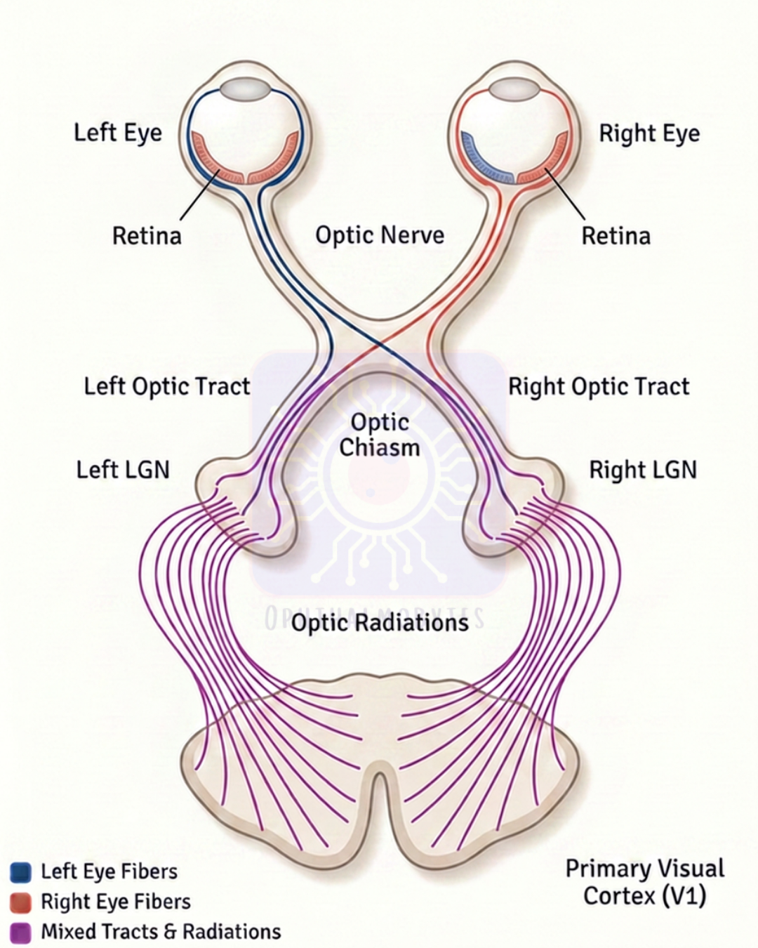
The afferent visual pathway begins in the retina and, as with other somatosensory pathways, has a three-order neuronal pathway, which ultimately relays information to the occipital cortex.
First-order neurons are formed by the bipolar cells of the retina, which synapse with retinal ganglion cells.
These second-order neurons extend their axons along the innermost portion of the retina, coalescing at the optic disc.
These axons exit the globe and continue posteriorly via the optic nerve, and both optic nerves join in the suprasellar region to form the optic chiasm.
The nasal retinal fibers (temporal visual field) decussate contralaterally, while the temporal retinal fibers (nasal visual field) remain ipsilateral.
The conjoined optic nerve fibers now form the optic tracts. The optic tract axons synapse in the lateral geniculate body (LGB) of the thalamus.
These, now third-order neurons, relay information from the LGB via the optic radiations (either the dorsal bundle or Meyer loop) to the visual cortex in the occipital lobe.
Image from Rajan Eye Care Hospital
#ophthalmology #ophthal #doctor #health #medical #vision #education #optometry #medicalstudent #optometrist #medicine #eye #ophtho #ophthalmologist #ophthalmo #med #medicaleducation #ophthalmologyresident #ophthalmologyresidency #apaoyo #anatomy #visualpathway #opticnerve #opticchiasm #optictract #opticradiations
